
Challenge
Immunohistochemistry (IHC) staining is routinely used for assessing antigen expression in tissues. However, manual analysis of co-expression and antigen interactions is cumbersome in monoplex IHC assays due to artifacts, staining variability and slice differences. Manual scoring of co-expression is also challenging because of the limitations to what the human eye can reliably detect and quantify.
The team at GenMab was interested in quantifying colocalization of immune cell markers and PD-L1 from H&E and sequential IHC slides to understand the spatial arrangement of immune cell classes with respect to tumors.
Solution
We employed a deep learning (DL)-based solution to align sequential monoplex IHCs, generating a virtual multiplex (VM) image and enabling co-expression analysis.
Sequential slides stained for H&E, CD3, CD8, CD163, and PD-L1 IHC were collected from 120 non-small-cell lung cancer (NSCLC) patients. Deep learning models were trained on each stain separately to classify tumor cells, lymphocytes, granulocytes and fibroblasts, and tumor versus stromal areas. To identify co-expression of IHC markers, the positive and negative density maps of each stain were interpolated at each H&E cell detection to determine its positivity or negativity for each marker.
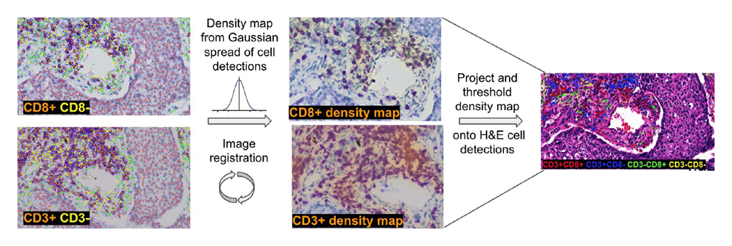
Figure 1. Workflow of the VM model generation
The resulting VM slide was generated using the AI-powered solution, and the colocalization image was assessed for sensitivity and specificity. To quantify sensitivity, we calculated the fraction of CD3+CD8+ (true positive) lymphocytes from CD3+CD8+ and CD3-CD8+ (false negative) lymphocytes, as all CD8+ lymphocytes should co-express CD3. Specificity was calculated as the proportion of CD8- non-lymphocytes (true negative) from CD8- and CD8+ (false positive) non-lymphocytes, since CD8 is expressed exclusively on lymphocytes (Figure 1). Sensitivity of the VM was 70% and specificity was 90%. IHC cell detection models reached an average accuracy of 81% across different cell types and IHC stains.
This method allows researchers to quantify co-expression between multiple proteins at single-cell resolution without the additional costs of developing a multiplex antibody assay.