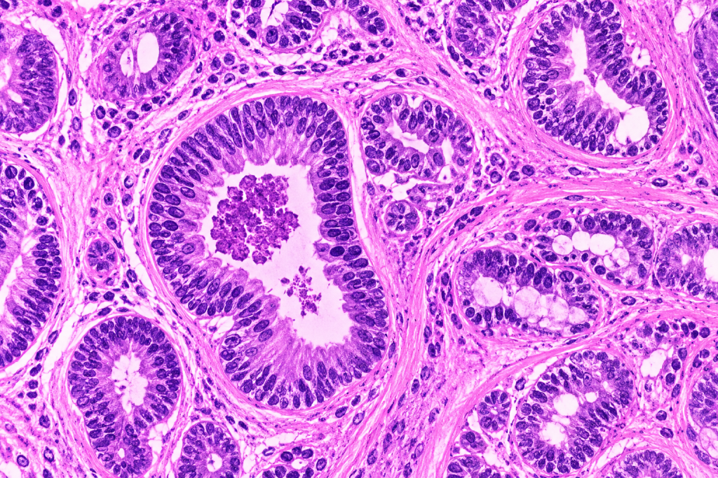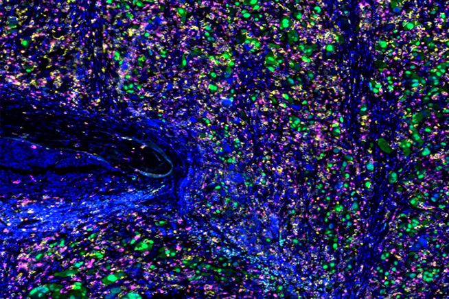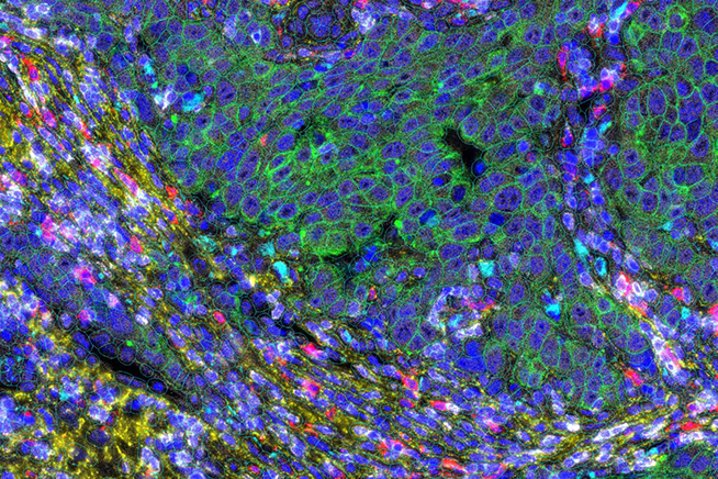

H&E, IHC, multiplex imaging, and spatial transcriptomics can each provide complementary views of tissue biology, and these data can be integrated to build a more complete picture of the tissue microenvironment.

AI-powered quantitative analysis produces structured, reproducible features that capture cellular composition, expression patterns, tissue architecture, and spatial relationships at a depth and consistency difficult to achieve through visual assessment or traditional computational tools.

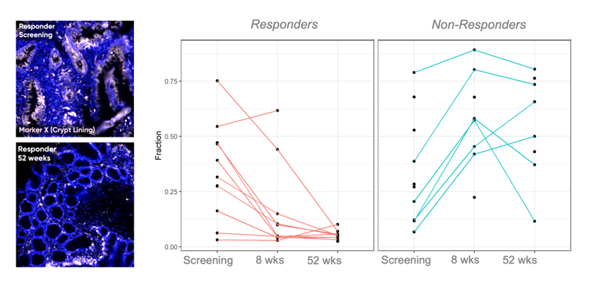
Tissue-derived features and thresholds can be systematically evaluated against clinical and outcomes data to establish which findings are associated with response, resistance, and survival.

Every finding can be traced back to the tissue image it came from, allowing scientists and pathologists to review the underlying biology and interpret what the quantitative result represents.