The search for better biomarkers of antibody-drug conjugate (ADC) efficacy just received a “hot tip:” new research reveals that cathepsin L (CTSL), a protease found in the tumor microenvironment, can enable ADCs to effectively target and kill HER2-low or HER2-negative cancer cells even when those cells do not express the HER2 target antigen (1). As Dr. Ken Bloom, Nuclei’s Head of Pathology, discussed in a recent webinar, this finding has profound implications for expanding the exploration of ADC mechanisms and biomarkers through AI-powered digital pathology.
Known ADC Mechanisms of Action: Antigen Targeting, Bystander Effects
ADCs combine a monoclonal antibody with a cytotoxic payload, connected by a carefully designed linker. The traditional mechanism of action for ADCs such as trastuzumab deruxtecan (T-DXd), with cleavable linkers, involves the antibody binding to its target antigen on the tumor cell surface. Thereafter, the entire ADC is internalized through endocytosis, and the payload is released within the acidic lysosomal environment to kill the cell.
In addition to direct killing of the cells expressing the tumor antigen on the surface, ADCs can also act via spatially-dependent bystander effects, where the payload diffuses out of the therapeutically-treated cancer cells and can suppress tumor progression in neighboring, untreated cells.
Whether ADCs act via direct antigen targeting or bystander effects, ADC pharmacology is complex, depending on binding, internalization, linker cleavage, cytotoxicity, and likely other processes. The case of two different HER2-targeted ADCs, trastuzumab emtansine (T-DM1) and T-Dxd illuminates just how complex ADC mechanisms can be.
Both the efficacy and resistance associated with T-DM1 in breast cancer are linked to HER2 expression levels and tumor heterogeneity, likely because T-DM1 lacks a cleavable linker and does not exhibit a bystander effect (2). In contrast, T-DXd, one of the most successful ADCs in clinical use, has a cleavable linker and a proven bystander effect, and shows efficacy across a broader range of HER2 expression levels—including high and low expressors (3,4).
The Cathepsin L Breakthrough: Is Target Antigen Expression Required for the Bystander Effect?
Recent research from teams at Duke University may have solved the mystery of T-DXd’s mechanism of action by demonstrating that T-DXd can achieve efficacy in HER2-low and even HER2-negative breast cancers through a mechanism that bypasses the requirements for target engagement and internalization (1).
The key player in this mechanism is cathepsin L (CTSL), a cysteine protease found in the tumor microenvironment. Unlike the traditional pathway that requires HER2 binding and cellular internalization, this alternative mechanism relies on extracellular proteases to cleave the ADC linker and release the cytotoxic payload directly into the tumor environment.
The research team compared T-DXd with trastuzumab emtansine (T-DM1), another HER2-targeting ADC with a non-cleavable linker. Using cell lines with high, low, and negative HER2 expression, they made several critical observations:
- Cytotoxicity Paradox: Despite poor internalization, T-DXd demonstrated significant cytotoxic activity against HER2-low and HER2-negative cells, while T-DM1 showed minimal activity.
- The Cathepsin L Connection: When the authors treated low-expressing cell lines with cathepsin L, an extracellular protease, they observed dramatic increases in T-DXd efficacy (Figure 1). Cathepsin L is significantly overexpressed in both the tumor and stroma of invasive breast cancers compared to normal tissue, and its expression is independent of HER2 status (Figure 2).
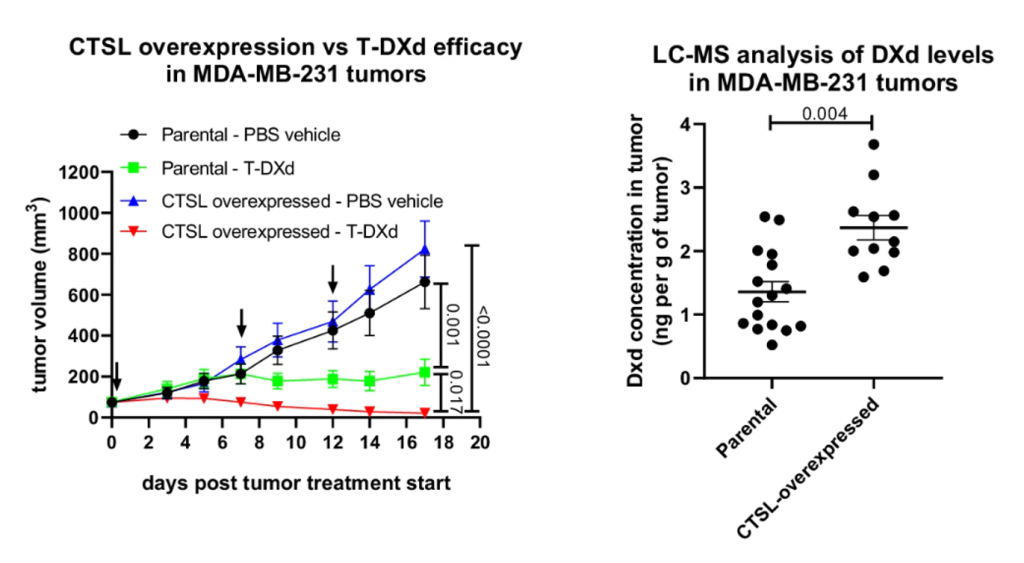
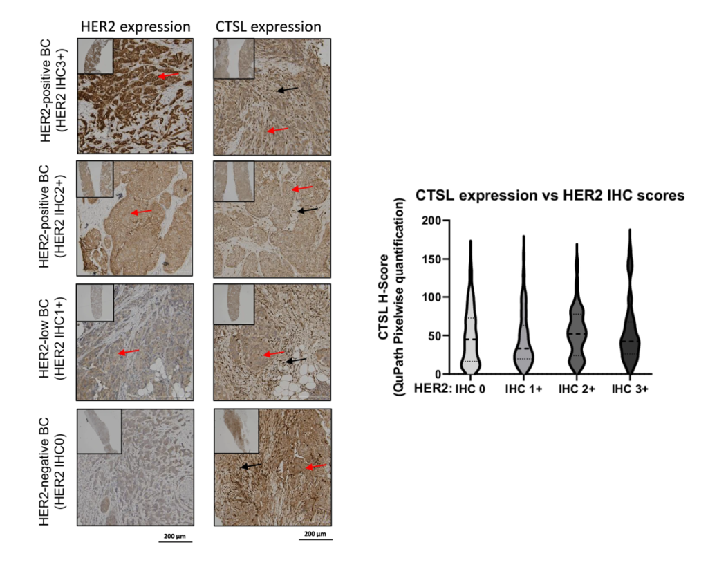
Figure 2. Cathepsin L is overexpressed in both the tumor and stroma of invasive breast cancers compared to normal tissue, and its expression is independent of HER2 status. Immunohistochemistry analysis of primary core needle biopsies from BC patients (left). Representative HER2 expression (left column) is shown. CTSL expression and localization in the same patient’s biopsy are shown (right column). Positive staining in tumors (red arrows) and stroma (black arrows) are indicated. Pixelwise H-scores were used for quantification by QuPath, with one-way ANOVA with Tukey’s multiple comparisons test, showing CTSL expression among BC with different HER2 IHC scores (right).
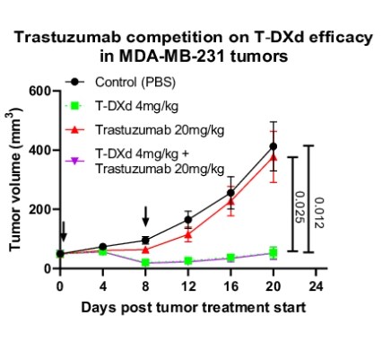
- Antigen-Independent Efficacy: When researchers administered excess trastuzumab to compete for HER2 binding sites, T-DXd remained effective against HER2-low cells, confirming that HER2 engagement is not necessary for therapeutic activity. (Figure 3)
Implications for Treatment Decisions and Therapeutic Development
This discovery has immediate implications for ADC design, drug development, and patient care. The cathepsin L-mediated mechanism may help explain why T-DXd has shown clinical efficacy across the spectrum of HER2 expression levels, including patients previously considered unsuitable for HER2-targeted therapy.
Cathepsin L expression is significantly higher in invasive breast cancer than normal breast tissue, and it is unrelated to HER2 expression levels or disease stage1. Therefore, cathepsin L expression could serve as a predictive biomarker for response to ADCs that have linkers cleavable by cathepsin-L , impacting clinical trial designs in breast cancer and potentially expanding treatment options for thousands of patients who were previously excluded from HER2-targeted therapy.
The Immunomodulatory Dimension
Beyond direct cytotoxic effects, the study revealed that T-DXd induces immunogenic cell death and activates key immune pathways, including STING and toll-like receptor 4 (TLR4) pathways1. This immunomodulatory activity opens new possibilities for combination therapies or multi-specific therapies employing checkpoint inhibitors and other immunotherapies, potentially achieving improved overall survival.
AI-Powered Digital Pathology and Spatial Proteomics: The Key to Unlocking Complex Mechanisms
The cathepsin L discovery likely represents the first of many discoveries that will shift in our understanding of ADC mechanisms, moving from target-based and bystander effect-based selection to comprehensive tumor ecosystem analysis. As more ADCs enter clinical development with diverse payloads and linker chemistries, the need for sophisticated biomarker strategies becomes even more critical.
Traditional pathology approaches, while valuable, struggle to capture the intricate relationships between multiple biomarkers, spatial distributions, and microenvironmental factors that influence therapeutic response.
This is where AI-powered digital pathology platforms become indispensable. Advanced computational pathology systems can now analyze tissue samples with unprecedented precision, measuring not just the presence or absence of biomarkers, but their spatial relationships, distribution patterns, and quantitative, continuous scoring of protein expression levels.
For ADC development, AI-powered digital pathology, in combination with multiplex spatial analysis, can simultaneously assess target expression, cathepsin L levels, immune cell infiltration, and other microenvironmental factors that influence therapeutic response. This technology can identify subtle patterns, missed by human pathologists, that might predict bystander effects. Also AI-powered digital pathology can measure tissue heterogeneity that affects drug distribution, and quantify spatial proximity scores that capture the complex interplay between different cell populations.
A Comprehensive Suite of ADC Solutions
At Nucleai, we’ve built a comprehensive ADC platform that can empower biopharma translational and biomarker teams to go beyond basic presence/absence measurements or manual H-score evaluations. Our platform goes further, delivering quantitative, optical density (OD)-based IHC quantification and spatial proximity analyses. This allows for a deeper understanding of mechanism of action, resistance, patient stratification, and therapeutic strategies that maximize benefit while minimizing toxicity.
Book a demo with us to discuss your research challenges, AI-augmented biomarker scoring, and the latest developments in our digital pathology platform.
References:
-
Tsao, LC., Wang, J.S., Ma, X. et al. Effective extracellular payload release and immunomodulatory interactions govern the therapeutic effect of trastuzumab deruxtecan (T-DXd). Nature Communications, 16, 3167, Apr. 2025. PubMed, https://doi.org/10.1038/s41467-025-58266-8.
-
Filho, Otto Metzger, et al. “Impact of HER2 Heterogeneity on Treatment Response of Early-Stage HER2-Positive Breast Cancer: Phase II Neoadjuvant Clinical Trial of T-DM1 Combined with Pertuzumab.” Cancer Discovery, vol. 11, no. 10, Oct. 2021, pp. 2474–87. PubMed, https://doi.org/10.1158/2159-8290.CD-20-1557.
-
Modi, Shanu, et al. “Trastuzumab Deruxtecan in Previously Treated HER2-Low Advanced Breast Cancer.” The New England Journal of Medicine, vol. 387, no. 1, July 2022, pp. 9–20. PubMed, https://doi.org/10.1056/NEJMoa2203690.
-
Bardia, Aditya, et al. “Trastuzumab Deruxtecan after Endocrine Therapy in Metastatic Breast Cancer.” The New England Journal of Medicine, vol. 391, no. 22, Dec. 2024, pp. 2110–22. PubMed, https://doi.org/10.1056/NEJMoa2407086.