

Why spatial biomarker strategy must scale for antibody–drug conjugates
The ADC paradox: precision drugs, fragmented biomarker strategy
Antibody–drug conjugates (ADCs) are often described as precision therapeutics. They are designed to exploit specific antigens, deliver potent payloads, and – in principle – limit off‑target toxicity. Yet in practice, ADC development continues to face a familiar paradox: heterogeneous clinical response despite apparently homogeneous target expression.
Traditional biomarker strategies—largely centered on average antigen expression or binary positivity thresholds—have proven insufficient to explain why some patients respond deeply while others do not. Spatial biology has begun to fill this gap, revealing how antigen distribution, tumor architecture, immune context, and cell–cell proximity shape ADC efficacy.
Why single‑trial spatial analysis falls short for ADC programs
Single trial spatial analyses provide important insights into the relationship between tumor architecture and ADC activity, but their limited scope can obscure broader, program level trends. Without integrating findings across multiple trials and cohorts, it becomes difficult to distinguish reproducible biological mechanisms from trial-specific artifacts. This challenge is heightened in ADC programs, where multiple indications, line of therapy settings, and companion biomarkers are often explored simultaneously. As a result, scaling spatial biomarker strategies beyond individual studies is essential to capture the complexity of ADC biology and ensure robust, generalizable insights that can inform future ADC development and clinical decision-making across diverse clinical contexts.
A visual framework: from trial insight to program strategy
To unlock the full value of spatial data in ADC development, analysis must intentionally scale beyond the individual trial.
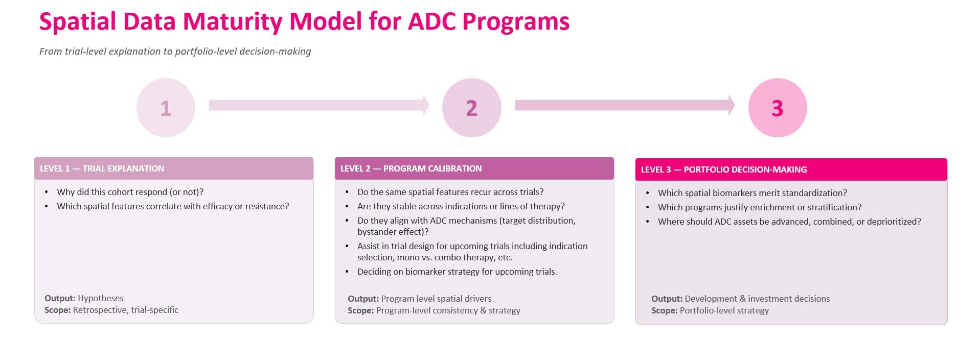
ADC‑specific insight: spatial context matters as much as target presence
ADCs are uniquely sensitive to spatial biology because their efficacy depends on physical relationships:
- Distance between target‑positive and target‑negative cells
- Clustering versus dispersion of antigen‑expressing tumor cells
- Proximity of immune or stromal components that influence payload diffusion
A biomarker strategy that focuses solely on average antigen expression can therefore miss critical determinants of response. Spatial analysis exposes these dimensions—but only repeated, cross‑trial observation can distinguish true biological drivers from trial‑specific artifacts.
For example, if spatial clustering of target‑positive cells correlates with response in one trial, program‑level analysis asks:
- Does this pattern recur in other indications treated with the same ADC?
- Does it persist across biopsy sites or disease stages?
- Is it consistent across different staining protocols or image analysis methods?
Only then can such a feature credibly inform future trial design or enrichment strategy.
Cross‑trial spatial validation: reducing false confidence
ADC development is particularly vulnerable to over‑interpreting early signals. Payload potency and narrow therapeutic windows amplify the consequences of biomarker miscalibration.
Program‑level spatial validation addresses this risk by:
- Testing whether candidate spatial biomarkers reproduce across studies
- Identifying features that are mechanistically coherent with ADC MoA
- Eliminating signals that fail to generalize beyond a single cohort
This mirrors the evolution of genomic biomarkers in oncology, which only became strategically useful once validated across multiple trials and contexts. Spatial biomarkers require the same discipline – especially given their higher dimensionality.
Prevalence and feasibility: the operational reality of spatial biomarkers
A spatial biomarker may be biologically compelling yet impractical at scale. For ADC programs, program‑level prevalence studies are essential to answer:
- How frequently does this spatial phenotype occur across the target population?
- Is prevalence sufficient to support enrichment or stratification?
- Does prevalence change with prior therapy, tumor evolution, or sampling site?
Without these answers, spatial biomarkers risk becoming scientific curiosities rather than developmental assets.
How program‑level spatial strategy shapes biomarker decisions in ADC development
By treating spatial analysis as a core resource across the program, teams can make informed biomarker decisions that guide trial enrichment and stratification strategies, especially in prospective settings.
- Biomarker strategy: selecting spatial features suitable for prospective enrichment or stratification, rather than exploratory post‑hoc. Nucleai biomarkers have shown improved effect size of close to x2, reducing screening by hundreds of patients which may result in tens of millions of dollars in per-trial savings.
- Indication prioritization: assessing prevalence of relevant spatial phenotypes to determine where an ADC is most likely to be effective
- Therapy context: distinguishing spatial drivers of response in monotherapy versus combination settings
- Development focus: advancing programs where spatial biology aligns with mechanism, while deprioritizing those where it does not
Ultimately, this approach leads to greater clarity in program‑level decisions, enabling more precise and impactful ADC development.
From trial‑level insight to program‑level biomarker strategy: Nucleai’s experience
Across ADC development, Nucleai has worked at the intersection of trial‑specific spatial analysis and program‑level decision‑making, supporting teams as insights evolve from explaining individual studies to informing forward‑looking biomarker strategy.
At the trial level, Nucleai has supported a wide range of clinical studies—from early phase through phase 3 – using spatial analysis of IHC, H&E, and multiplex immunofluorescence (mIF). These analyses include ADC‑specific features related to target distribution, tumor architecture, immune context, OD-based compartment intensity, and spatial proximity, enabling identification of biomarkers associated with response or resistance within individual cohorts.
A clear example of going into a program level comes from Nucleai’s work in small cell lung cancer (SCLC). In this setting, Nucleai developed a dedicated H&E‑based SCLC algorithm, trained once using a duplex stain as a reference to establish accurate cellular and structural ground truth (1). This approach enabled development of a model that captures disease‑specific morphology and spatial context with a level of consistency that would be difficult to achieve using H&E review alone. Importantly, the resulting algorithm is not tied to a single trial. Once established, it can be applied across real‑world datasets, completed studies, and ongoing clinical trials within the same program, creating a shared analytical foundation.
Nucleai has also been involved in program‑level ADC efforts, where spatial insights were carried forward across studies. In these settings, Nucleai deployed prospective digital pathology AI algorithms within specific clinical trials and complemented them with prevalence studies, cell line analyses, and additional spatial scoring. Together, these efforts supported decisions around indication selection, biomarker feasibility, and broader program direction.
Nucleai analyzed multiple phase 3 trials within the same ADC programs, enabling comparison of spatial biomarkers across trials rather than in isolation. This work yielded both trial‑specific biomarkers and program‑level insights, including the recurring importance of H&E‑derived spatial features alongside IHC biomarkers in understanding ADC efficacy.
At the single‑trial level, spatial biomarkers have been shown to meaningfully amplify treatment effects (2,3) when used for enrichment or stratification. At the program level, however, the added value comes from understanding how these biomarkers change across trials and indications, and how they differ between monotherapy and combination settings. Notably, the addition of immuno‑oncology agents to ADCs can significantly alter the spatial biomarkers most relevant to response, underscoring the need for program‑level calibration rather than one‑time discovery.
Taken together, this experience highlights a central lesson: spatial biomarkers are most powerful when treated not as isolated trial readouts, but as shared program assets—continuously tested, refined, and aligned with mechanism as development progresses.
Looking forward: spatial biology as an infrastructure
The next phase of ADC development will not be defined solely by better linkers or payloads. It will be shaped by how well organizations integrate spatial understanding into program‑level decision‑making.
Spatial analysis should no longer answer only:
- “Why did this trial behave the way it did?”
But instead:
- “What does this teach us about how to design the next five trials?”
For ADC portfolios, that shift may be the difference between incremental progress and durable precision.
Connect with Our Team
Interested in learning more? Fill out the form below to connect with our team
and explore how Nucleai can support your biomarker and clinical development goals.