

A representative image of a responder PPP low and a non-responder PPP high demonstrating differences in ASCT2 (a glutamine transporter) and G6PD and pNRF2 (regulators of the pentose phosphatase pathway)
Checkpoint inhibitor immunotherapies have broadened treatment possibilities for cancers like lung cancer, yet only 15-30% of lung cancer patients see positive responses. For the remaining majority who don’t experience clinical benefit, a recent study led by Dr. Arutha Kulasinghe, in collaboration with the scientific team at Nucleai, may offer a promising solution.
Dr. Kulasinghe’s work represents the first study investigating the role of metabolic signaling-based biomarkers in immunotherapy-treated lung cancer patients.
Connecting tumor cell metabolism to lung cancer immunotherapy response
Dr. Kulasinghe’s team at the Frazer Institute, University of Queensland, Australia employed Nucleai’s AI multiplex image analysis solution to explore hallmarks of cancer that were associated with clinical endpoints of immunotherapy treatments.
One well-known cancer hallmark, the increased glucose consumption by tumor cells known as the Warburg effect, may also modulate the response of immune cells in the tumor microenvironment (TME) — by depleting the TME of nutrients. Studies in melanoma and breast cancer reveal metabolic signatures associated with immunotherapy resistance.
What tumor hallmarks could be enabling immunotherapy resistance in non-small cell lung cancer (NSCLC)?
Using samples from NSCLC patients, Dr. Kulasinghe’s team performed multiplex immunofluorescence (mIF) using the PhenoCycler(R) Fusion platform from Akoya Biosciences with a 45-plex protein biomarker assay panel covering tumor, immune and metabolic content.
The team discovered a subset of tumor cells with upregulated proteins in the pentose phosphate metabolic pathway (PPP), which was characterized by upregulation of ASCT2, a glutamine transporter, as well as pNRF2 and G6PD. These “PPP high” cells displayed characteristics of aggressive tumor cells, such as higher proliferation and lower differentiation. These cells also showed lower immunotherapy response rate and lower survival (Figure 1).
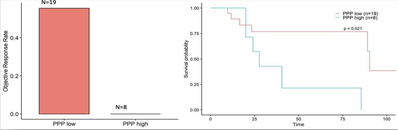
Figure 1. Novel tumor cell subtype with high metabolic activity shows lower immunotherapy response rate and lower survival.
“The study’s findings shed light on how the functional and metabolic activity of tumor cells causes them to proliferate aggressively and develop resistance to immunotherapy,” explained Dr. Kulasinghe. “Our study has unveiled a unique metabolic signature in tumor cells that is closely linked to immunotherapy resistance in NSCLC cancer patients.”
The Nucleai solution: AI-based spatial biomarker analysis for unprecedented accuracy
Nucleai’s platform is the first commercially available solution for deep learning-based multiplex image analysis.
Traditional multiplex image analysis involves quantifying mean marker intensity on segmented cells, but this quantitation can be inaccurate and imprecise because of background noise, inconsistent segmentation, and imaging artifacts. Low-quality marker quantitation, when used with manual gating and traditional clustering-based cell typing methods, leads to inaccurate cell typing.
The Nucleai solution, going beyond marker intensity and traditional cell segmentation, employs a more accurate, neural network approach to cell typing decisions, based on machine learning. Informed by millions of annotations, the Nucleai solution applies image quality control, then identifies tissue area segments, classifies cells, and quantifies spatial features, such as cell arrangements, neighborhoods, and tertiary lymphoid structures. Using this spatial context of surrounding features yields a more accurate way to quantify marker expression and identify cell subtypes.
Combining supervised cell typing with unsupervised clustering
In this study, the Nucleai team worked with Dr. Kulasinghe’s data to combine supervised cell typing with unsupervised approaches for identification of cell functional states. This methodology allowed for maintaining cell typing accuracy while identifying novel cell states which could be linked to clinical outcome.
The team performed unsupervised clustering of the expression vectors of metabolic and cell state proteins to further segment cells into functional subtypes (Figure 2). This unbiased, AI-enabled approach surfaced markers defining novel cell subtypes that would otherwise remain hidden.
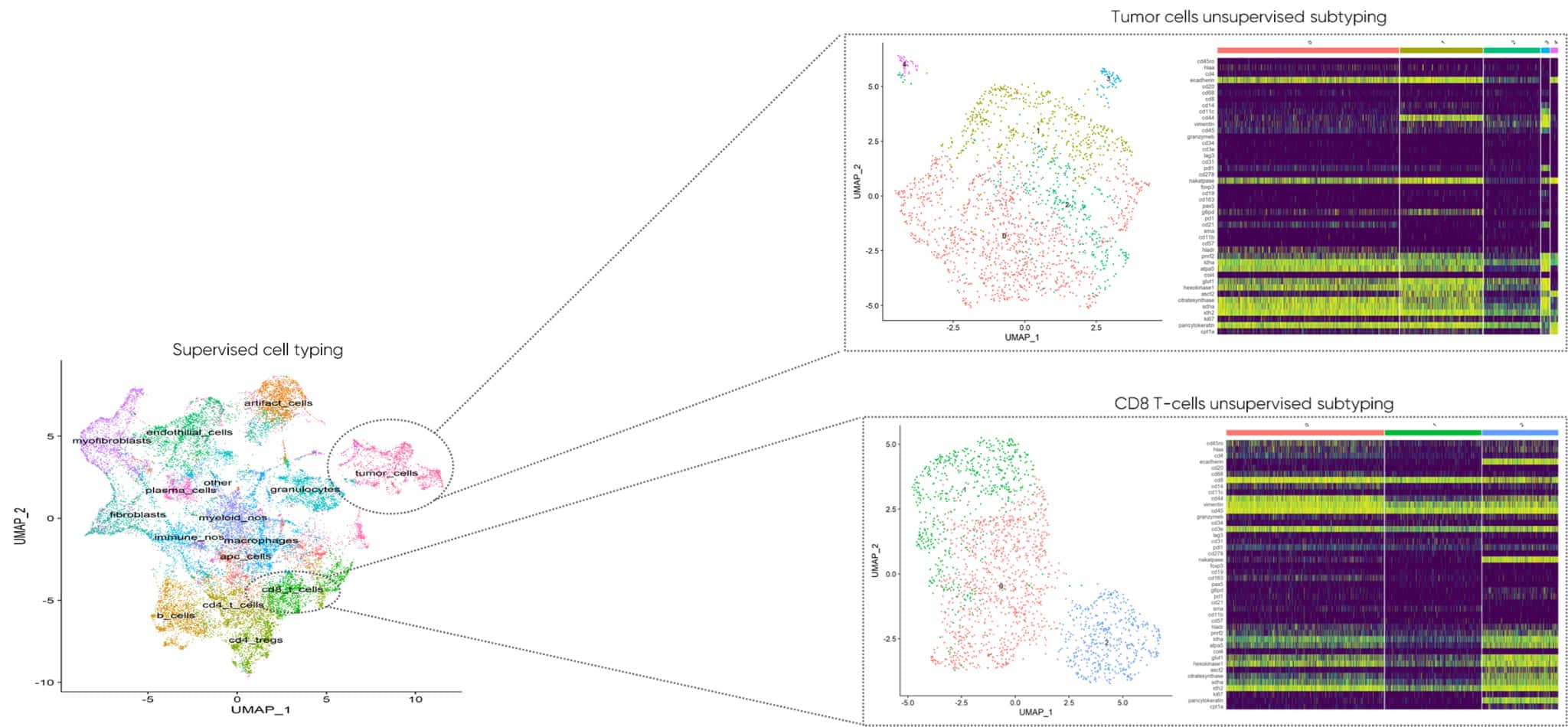
Figure 2. Unsupervised clustering of metabolic and cell state protein expression revealed functional signatures of tumor and immune cell subtypes.
“I think that’s the biggest takeaway,” said Dr. Kulasinghe, “We’re moving away from cell phenotypes, into functional annotation of these cell types, to determine which functional profiles are associated with clinical endpoints.”
Combined treatments targeting metabolism and immune checkpoints: the promise of overcoming resistance
Both Dr. Kulasinghe and the Nucleai team are excited by the potential for the results of this study (and subsequent validation studies) to increase treatment options for lung cancer patients.
By screening patients for the newly identified metabolic biomarker signature, clinicians could identify a subset of non-responders who may benefit from a combination approach. For example, these patients could receive metabolic inhibitors alongside the immune checkpoint inhibitor, potentially overcoming resistance mechanisms and increasing their likelihood of responding to immunotherapy. This personalized, biomarker-guided strategy could extend the reach of immunotherapies to a broader population of lung cancer patients.
This discovery holds the promise of improving immunotherapy response rates by providing crucial insights into disease progression, treatment response, resistance mechanisms, and the development of more effective therapeutics and diagnostics.
As we celebrate Cancer Immunotherapy Month, we hope that this study launches a new era in our understanding of cancer metabolism and its implications for personalized medicine.