AstraZeneca pioneers a novel AI biomarker for their ADC trials, a trispecific trial adopts AI spatial scoring, researchers publish the first extensive tumor maps, and a groundbreaking research-to-cure spatial discovery saves patient lives.
At the core of the evolving field of precision spatial medicine is a powerful idea: each patient’s biopsy contains the blueprint for guiding the right treatment decision. If this biological instruction manual can be properly decoded, the best treatment decision will come into focus.
Traditional diagnostic methods, often limited to binary or reductionist approaches, fall short in capturing the full complexity of patient biology. The interplay of cells and proteins, rather than simple gene mutations alone, is critical for understanding how to target diseases with an ever-expanding toolbox of advanced therapeutic options.
AI and spatial technologies have emerged as game-changers, unveiling the hidden biology and cellular interactions within each patient’s biopsy. These innovations are making AI-enabled spatial precision medicine a reality, paving the way for truly personalized treatments with advanced therapies and transforming how we understand and combat disease.
Breaking New Ground in 2024
Undoubtedly, 2024 has been a watershed moment for AI-enabled spatial precision medicine. The underlying technologies continued to advance rapidly, with seemingly limitless applications in oncology and beyond. Below, I’ve highlighted just a few milestones from the year that exemplify both the progress made and the immense promise of this approach. These achievements range from AI-enabled biomarkers for antibody-drug conjugates (ADCs) and bispecific trials, to a remarkable journey from discovery to cure that happened in just a few months. We’ll encounter the “method of the year” and see how AI-enabled technologies are tracking the evolution of tumors in space and time.
Let’s start the countdown.
5. Spatial Proteomics Named Method of the Year
Nature Methods named spatial proteomics the 2024 Method of the Year due to its role in uncovering the organization of complex tissues and its ability to produce highly detailed tissue maps that provide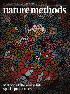 insights into protein composition and spatial arrangement of different cell types.
In selecting spatial proteomics, the editors noted their excitement as the field further evolves new ways to explore the spatial proteome in greater depth and breadth. Efforts of large consortia such as the Human BioMolecular Atlas Program (HuBMAP) and the Human Tumor Atlas Network (HTAN) are lauded, along with advancements in AI-based methods to process, analyze, visualize, and mine spatial data.
Being named Nature’s Method of the Year is a prestigious recognition as the title is awarded to a method that has shown transformative promise in advancing research or clinical applications. This distinction shines a spotlight on groundbreaking innovations that can dramatically shift the course of medical progress. For example, Nature’s 2007 Method of the Year was next-generation sequencing (NGS), which, within a few years, became a critical enabler of targeted therapies, dramatically changing the landscape of precision medicine. Such recognitions often serve as a catalyst for further investment and development, helping drive progress in the field.
4. HTAN Publishes Seminal Studies Tracking Tumor Evolution in Space and Time
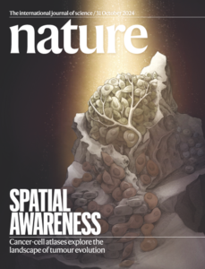
Founded in 2018 and consisting of ten research centers, HTAN, a US National Cancer Institute (NCI)-funded Cancer MoonshotSM initiative, compiles atlases that integrate cellular, molecular, and histological features of tumors as they evolve. The goal of the atlases is to detail the biological processes that underlie the ability of cancer to initiate, progress, and either respond or resist therapy. In October 2024, Nature published a dozen new papers from HTAN, across 20 indications, several of which used spatial omics in their studies to show how cancer cells behave and interact over time and reveal new findings about tumor evolution. Several of these studies were highlighted by Dr. Eric Topol, a globally renowned physician, including one that demonstrated that colon cancer often starts with multiple clones of cells that cooperate, before one ultimately dominates at the pre-cancerous stage. Another study mapped the immune response in breast cancer and identified hot zones of high immune activity where the immune system is attacking the tumor, and cold zones, where the tumor is evading the immune system. Such detailed spatial maps of tumor biopsies can guide combination immunotherapy strategies to tackle the complex neighborhoods in the tissue. Topol’s article also highlighted the impressive progress being made in cancer biology in the areas of diagnostics and treatments. He also emphasized the potential of AI-driven technologies, which are helping to crunch through colossal amounts of biological data, identify novel drug candidates, and predict the efficacy of treatments more quickly than traditional methods.
3. AstraZeneca Adopts a Novel AI-enabled Spatial Biomarker in a Phase III ADC-Combo Trial
The field of ADCs is experiencing rapid growth, with over 200 trials initiated in the past two years alone, signaling a significant opportunity for greater precision in cancer treatment. While most of the currently approved ADCs do not require biomarker testing, there is a pressing need for better predictive biomarkers. AstraZeneca recently outlined its strategy for using an AI-enabled biomarker to derisk the development of their Daiichi Sankyo-partnered datopotamab deruxtecan (Dato-DXd), a TROP2-directed ADC, in non-small lung cancer (NSCLC) patients. Trophoblast cell surface antigen 2 (TROP2) is a cell surface protein overexpressed in several types of cancer, and TROP2-directed ADCs have demonstrated potential in improving outcomes for patients with various cancers. The development of a predictive biomarker for TROP2 expression is critical to optimizing the use of this ADC, as it helps identify patients who are most likely to benefit from treatment.
AI-enabled biomarkers play a crucial role in addressing this unmet need by helping to identify the right patients for specific ADC therapies, predict resistance, and ultimately de-risk drug development efforts. These advanced tools leverage vast amounts of clinical data and computational methods to uncover patterns in tumor biology, enabling more accurate decision-making and better treatment outcomes.
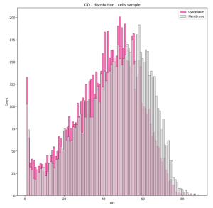
AstraZeneca has developed a proprietary, AI-powered biomarker algorithm that uses spatial information to measure the ratio of TROP2 expression on the cell membrane relative to expression in the cytoplasm of tumor cells, reflecting the importance of where the protein is expressed. This innovative approach uses Quantitative Continuous Scoring (QCS), a novel AI-enabled computational method for analyzing a pathology slide, which differs from traditional, semi-quantitative methods where a pathologist manually scores the slide with their naked eyes, without any assistance from deep learning algorithms. Their studies demonstrated that the spatial location of the protein expression is relevant to the biomarker’s predictive potential and the exact cutoff scores were optimized through a retrospective analysis of their phase 3 TROPION-Lung01 trial samples.
The biomarker is now being utilized in a couple of phase 3 trials evaluating Dato-Dxd in combination with other treatment modalities like immunotherapy, a bispecific antibody, and chemotherapy (AVANZAR, TROPION-Lung10).
2. A Leading Bispecific Biotech Adopts AI-aided Spatial Scoring for their Clinical Trial
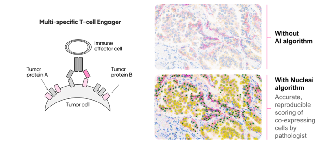
Multispecific antibodies can deliver improved targeting and efficacy by simultaneously binding multiple antigens. Identifying patients likely to respond to these advanced therapeutics requires evaluation of their tumor cells and immune cells to determine whether they co-express the corresponding antigens.
We recently collaborated with a top 10 biopharma company that sought to deploy an immunohistochemistry (IHC) assay for this purpose, and enroll patients in a Phase I trial of a multispecific drug. Pathologists had difficulty with manual scoring as it required accurate and reproducible quantification of tumor cells co-expressing the two target proteins. An AI-powered multiplex IHC (mIHC) algorithm that enables spatial scoring of co-expression and colocalization was developed, optimized, and validated. The AI-enabled assay was then deployed in partnership with the biopharma company and a leading in vitro diagnostic (IVD) company to aid pathologists with scoring co-expression and supporting the Phase I trial.
These trials underscore a broader trend of AI-driven precision medicine in oncology, where biomarker identification and patient stratification are key to maximizing treatment efficacy and improving patient outcomes.
1. AI-Enabled Spatial Discovery Makes Its First Impact by Saving Patient Lives
Scientific breakthroughs typically require decades of effort to move from the bench to the bedside – and that’s just the ones that make it across the finish line.
How about if we do it in less than a year? Researchers have published a groundbreaking discovery for treatment of toxic epidermal necrolysis (TEN), a severe, often fatal allergic reaction to certain medications that causes the skin to blister and peel. They used a spatial technique called deep visual proteomics (DVP), a combination of AI-driven analysis, microscopy and protein-analysis, to analyze skin biopsies in a way that retains spatial context information which can be used to create detailed spatial protein maps.
With this approach, a drug target in a hyperactive JAK/STAT signaling pathway in TEN was identified, a true needle in a very broad and very deep haystack of more than 5000+ proteins across thousands of cells.
This finding pointed to JAK inhibitors, already used for other inflammatory conditions, as a potential treatment. Repurposing this existing drug, the team tested it in vitro, in mouse models, and then in seven human patients, all of whom made a full recovery.
This breakthrough highlights the critical importance of understanding disease at the molecular level and the transformative potential of spatial medicine in developing targeted, life-saving therapies.
The significance of this success is best captured by Matthias Mann, lead author of the paper: “To our knowledge, this is the first time a spatial omics technology has made an immediate and tangible impact in the clinic, by identifying a treatment that has already changed people’s lives for the good. This approach could be applied to a wide range of diseases, potentially accelerating drug discovery across multiple fields of medicine.”
What Lies Ahead
Decades ago, our understanding of the difference between cancer and normal cells was relatively straightforward: cancer cells divide more rapidly. This insight fueled the development of early chemotherapies, designed to disrupt the processes most active in rapidly dividing cells.
Today, the complex tapestry of cancer cells and the tumor microenvironment continues to be revealed, and the hallmarks of tumor biology extend well beyond relatively simplistic distinctions. A vast array of cancer cell characteristics and vulnerabilities now form the foundation of next-generation therapies, such as ADCs, bispecific antibodies, and immuno-oncology (IO) combinations. These therapies employ sophisticated targeting to home in on cancer cells with unprecedented precision, which means their success relies on next-generation diagnostics – and this is where artificial intelligence (AI) and spatial technologies will play a pivotal role.
AI-guided spatial mapping of tissue biopsies, combined with advanced biomarker analysis, promises to revolutionize treatment decisions. By integrating and processing terabytes of data from imaging techniques like hematoxylin and eosin (H&E) staining, IHC, mIHC, and multiplex immunofluorescence (mIF), we can enhance biomarker scoring, better understand mechanisms of action (MOA), and predict therapeutic responses with greater accuracy.
At Nucleai, we’re proud to be at the forefront of AI spatial precision medicine and bringing innovative approaches to our customers as they bring disease biology into sharper focus and deliver new treatment options that will impact the quality and length of patients’ lives.
I’m excited to see what 2025 holds for this field, and I hope you are as well.